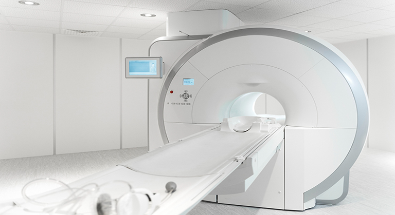
What We See Is What We Get
We've invested in a new way of looking into your body - a procedure that's fast, comfortable, and incredibly precise. Using digital radiography, we can clearly identify all external and internal anatomical structures and accurately diagnose your chest and bony problems. Even more amazing, we can immediately translate that information into a large, clear, accurate image, projected onto a monitor that patient and doctor can study together in the operatory. Digital radiography's technology improves and simplifies the way we care for our patient’s, resulting in better dental evaluations and treatment decisions.
Reduced Radiation, Radical Results
Traditionally X-rays are taken to see what the naked eye could not; X-rays were developed in a darkroom with hazardous chemicals, and then viewed on a special light board. The developed X-rays had to be stored, which required large filing systems. By far, the worst part of traditional X-rays was the radiation exposure to patients. Digital radiography has completely transformed this process. We can rotate and magnify it, adjust it for contrast, and even color-code it for educational purposes. The digital images store easily and efficiently in our computer files, safe and sound. For insurance purposes, referrals, or patient education, digital X-rays can be easily, inexpensively, and accurately reproduced.
Digital X-rays offer unparalleled benefits over traditional radiographs: they're convenient, safe for the environment, provide a great opportunity for patient education, can be transferred and copied accurately, and best of all, they're safer for our patients.