Abdominal & Renal Doppler
The liver, gall bladder, pancreas, aorta (Large artery that carries blood from heart to the different parts of body), spleen, biliary tree (bile and gallbladder ducts) or inferior vena cava (IVC - Large vein that returns blood to the heart from parts of the body below the diaphragm renal arteries : Arteries that supply oxgyented blood to the kidney on eithers sides) may be examined during an abdominal ultrasound.
Carotid & Vertebral Doppler
Carotid doppler ultrasound is a non-invasive test that uses sound waves to measure the flow of blood through the large carotid arteries that supply blood to the brain.
These arteries can become narrowed due to arteriosclerosis or other causes, and this can lead to transient ischemic attack (mini-stroke) or cerebral vascular accident (stroke). The carotid doppler test can help doctors determine stroke risk and the need for preventive measures.
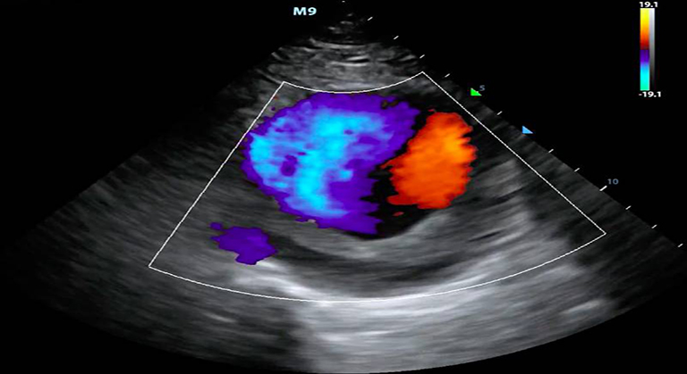
2 - D Colour Doppler Echocardiography
An echocardiogram (also called an echo) is a type of ultrasound test that uses high-pitched sound waves that are sent through a device called a transducer. The device picks up echoes of the sound waves as they bounce off the different parts of your heart. These echoes are turned into moving pictures of your heart that can be seen on a video screen.
Transthoracic echocardiogram (TTE). This is the most common type. Views of the heart are obtained by moving the transducer to different locations on your chest or abdominal wall. Stress echocardiogram. During this test, an echocardiogram is done both before and after your heart is stressed either by having you exercise or by injecting a medicine that makes your heart beat harder and faster. A stress echocardiogram is usually done to find out if you might have decreased blood flow to your heart (coronary artery disease).
Varicocele Doppler, Penile Colour Doppler
Varicoceles are abnormal dilatations of the pampiniform venous plexus. They are classified as primary or secondary, depending on their cause, and staged clinically on the basis of their extension and on the presence or the absence of spontaneous or induced reversal of blood flow.
Fetal Colour Doppler
Fetal middle cerebral arterial (MCA) Doppler assessment is an important part of assessing fetal cardiovascular distress, fetal anaemia or fetal hypoxia. In the appropriate situation it is a very useful adjunct to umbilical artery Doppler assessment. It is also used in the additional work up of
iintra-uterine growth restriction (IUGR) twin to twin transfusion syndrome (TTTS) twin anaemia polycythaemia sequence (TAPS)
Fetal echo blood of experiance advanced it requires technology machine to do complex sonography like fetal echo is detail evoluation of fetal heart in pregnant women at around 22 weeks of pregnancy. Ductus venosus spectral assesment is equally important for assesing fetal cardiovascular distress.
Peripheral Arterial & Venous Doppler
Doppler ultrasound is a technique used to measure the flow of blood through your arteries and blood vessels-usually those in your extremities. Vascular flow studies, also known as blood flow studies, can detect abnormal flow within a blood vessel. This can help to diagnose and treat a variety of conditions, including blood clots and poor circulation. A Doppler ultrasound can be used as part of a blood flow study.
This risk-free and pain-free procedure requires little preparation and can provide your doctor with valuable information about any for you arterial blockages you may have.
This risk-free and pain-free procedure requires little preparation and can provide your doctor with valuable information about any for you arterial blockages you may have.
Sonography guided Interventions
Interventional radiology is experiencing a period of unprecedented growth. Advances in equipment technology have facilitated the development of new treatment options and allowed refinements in existing techniques that have made established procedures safer.
Ultrasound is at the forefront of this trend, because it is becoming increasingly recognized as the premiere guidance tool for an array of interventional techniques. This article updates the radiologist on recent trends in ultrasound intervention and discusses several of the most significant ultrasound-guided interventional procedures at the turn of the twenty-first century.